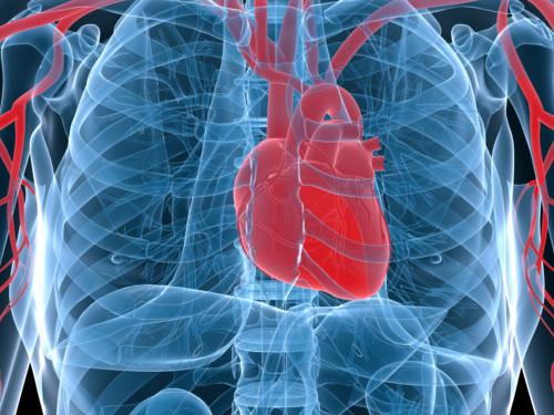
The reactive heart
Researchers look to heart muscle cells to understand what makes the heart tick
Published online 24 December 2014
The heart is constantly regulating its pulse rate by small changes in proteins present in the muscle cells
© Sebastian Kaulitzki / Alamy
Researchers are taking a closer look at the actual heart muscle cells that are responsible for pumping blood around the body, hoping to unlock new treatments for some of the most widespread chronic diseases.
Your heart is a mechanical organ that beats more than 100,000 times per day. Far from being a simple pump under exclusive control of the autonomic nervous system, the heart itself can detect changes in the demands placed upon it, and adjust its actions accordingly on a beat-by-beat basis.
This auto-regulation is possible, in part, thanks to mechanically-sensitive proteins embedded in the membranes of heart muscle cells, including stretch-activated ion channels (SACs) that respond to membrane deformations to alter the flow of electrical charges across the membrane.
SACs were first identified about 30 years ago in heart muscle cells isolated from chick embryos. Although they probably mediate the heart’s mechanical sensitivity, researchers have yet to identify them in the main heart chambers of mammals and have little understanding of how they work.
A recent review, published in Global Cardiology Science & Practice, summarizes what is known about heart SACs and potential future developments that may have clinical relevance1.
“A lot remains to be discovered in this exciting field,” says senior author Rémi Peyronnet, from Imperial College London, United Kingdom. “We still don’t know what tensions are present where in a cell, or exactly what activates the channels in vivo.”
Researchers have identified a number of molecules that exhibit the appropriate electrophysiological properties of SACs, with transient receptor potential (TRP) channels, which are widely expressed in several organs, being among the most likely candidates.
Several TRP channels are found in the heart. For example, antibody staining experiments in rat heart muscle cells show that the distribution of TRPC6 is consistent with a role in sensing membrane stretching. Some studies further demonstrate that it is sensitive to mechanical forces, but others provide conflicting results regarding both its sensitivity and its distribution.
Other candidates include several different types of potassium channels. One of these is TREK-1, which is regulated by membrane deformation and stretch, as well as other types of stimuli. Another is a channel called KATP, which exhibits mechanical sensitivity and is expressed in the heart after infarction and other diseases.
Stretch-activated channels are implicated in a wide range of diseases, including depression and muscular dystrophy, says Peyronnet. “[They] are obvious therapeutic targets. Channel- and organ-specific inhibitors may be of significant utility for improving patient conditions, and [some of these] are already under investigation.”
Reference
- Reed, A., Kohl, P. &Peyronne, R. Molecular candidates for cardiac stretch-activated ion channels. Glob. Cardiol. Sci. Pract. 2014 (2), 19 (2014). | article
DOI: 10.1038/qsh.2015.41
